- +91 417 922 0553
- office@shctpt.edu
- Contact Us
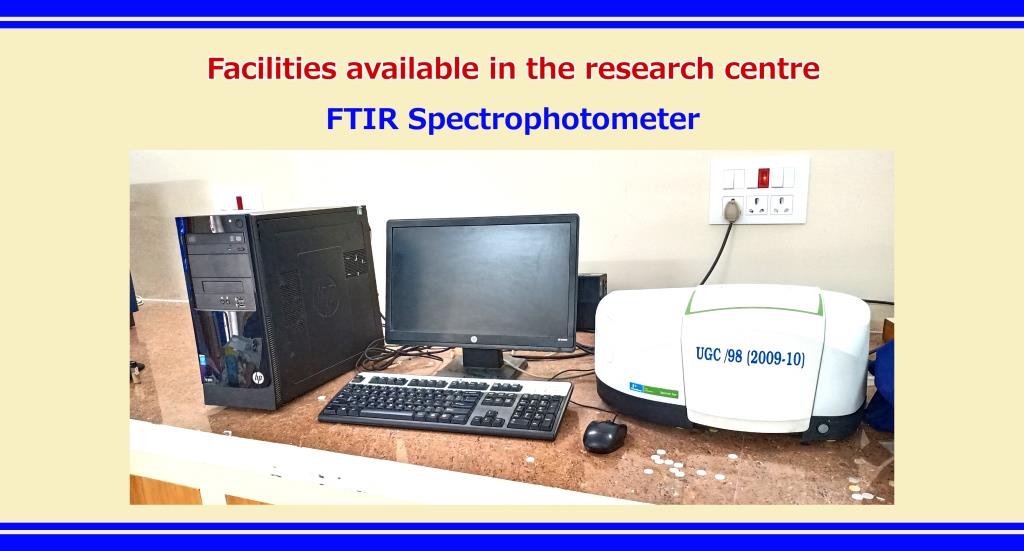
FTIR spectrophotometer is used for the identification of unknown compounds,
quantitative information, such as additives or contaminants, the identification of
functional groups of organic compounds, polymer materials, petrochemical products, biological compounds,
the pharmaceutical products and for academic research
Technical Description and Major Specifications
Model : PerkinElmer spectrum two FTIR spectrometer
Wavenumber range : 4000 to 350 cm-1
Detector : TGS detector
Resolution : 0.5 cm-1
S/N ratio : 2000:1 ppm for 1 minute scan.
Wavelength accuracy : 0.01 cm-1
Sample requirement :Solid: ∼0.2 (powder)
Charges:
Internal samples: Rs.50/- per sample
Outside samples: Rs.100/- per sample
Click Here to Download Sample submission form for FTIR spectrometer.
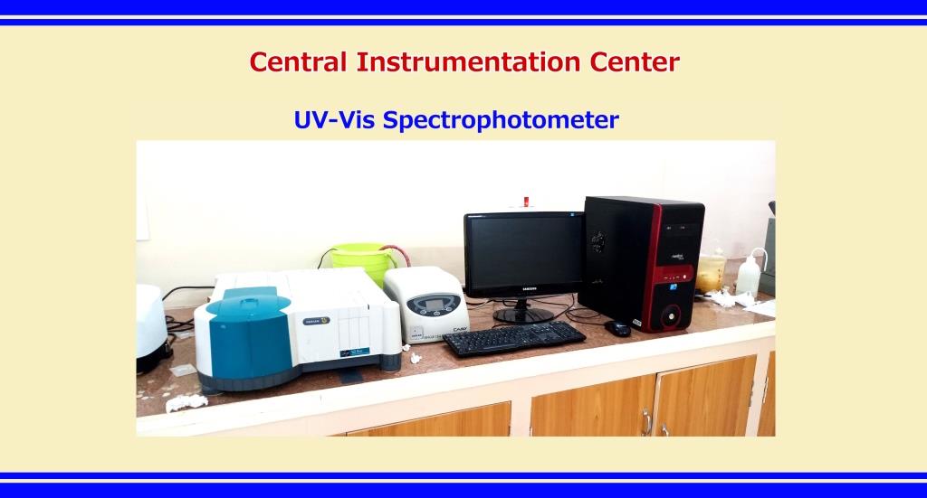
The Varian Cary 50 Scan UV Visible Spectrophotometer is designed to analyze microlitre volumes of
chemical and biological samples. With a maximum scan rate of 24,000 nanometers per minute,
the Varian Cary 50 can scan the whole wavelength range of 190 to 1100 nm in less than 3 seconds.
Additionally, the Varian Cary 50 features a data collection rate of 80 points per seconds and can measure samples
up to 3 Abs to reduce the need for frequent dilution. The Varian Cary 50’s super-concentrated beam makes it ideal
for fiber optic work, offering excellent coupling efficiency.
Technical Description and Major Specifications:
Model : Cary 50 UV
Source : Deuterium lamp (UV region)
Beam splitting system : Beam splitter
Detectors : Bandwidth dual Detector
UV-Vis limiting resolution : ≤ 1.5 nm
Wavelength range : 190 to 1100 nm
Photometric range : 3.3 abs
Cell Holder : Single cell holder
Resolution : ≥ 1.65 nm (UV/Vis)
Spectral bandwidth : Fixed at 1.5 nm
Maximum scan rate : 24,000 nm/min
Dimensions (W x D x H) : 50 x 59 x 20.5 cm
Data acquisition modes : Spectrum, kinetics and photometric with facility such as % Abs, %T, %R, absolute %R, log Abs, 1st-4th derivative
Note:
Instructions for UV-VIS users:
• Please specify the mode of measurement (% A,%T,%R etc)
• Solvents must be specified for recording solution spectra
Sample requirement:
• Solid: ∼0.2 (powder)
• Thin film: 1.0-1.5 cm
• Solution: ∼5 ml
Click Here to Download Sample submission form for UV-Vis spectrometer.
General Information
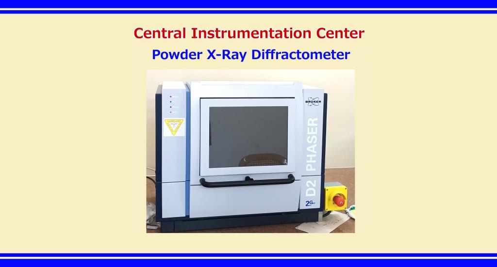
Model : Bruker D2-Phaser
Year of Installation : 2018
Specification
X-Ray Source : 2.2kW Cu anode, 40 kV/40 mA
Detector : LINXEYE XE
Step size : 0.0002o
2Theta range : 4 o to 80o
About Instrument and its applications
X-ray Diffraction (XRD) is a high-tech, non-destructive technique for analyzing a wide range of materials,
including metals, polymers, catalysts, plastics, pharmaceuticals, thin-film coatings, ceramics, s
olar cells and semiconductors. The Bruker D2 X-ray diffractometers can be used for nearly all X-ray diffraction application,
such as structure determination, phase analysis, stress and texture measurement. The D2 PHASER delivers good data quality and due to its compact size,
low weight, and ease-of-use design, the system is conveniently mobile, without the need for complicated infrastructure. A standard power outlet
and a few minutes is required to take for the system for an outstanding results. The D2 PHASER is a well equipped instrument with the unique LYNXEYE XE-T detector,
energy resolution of 380 eV allows a unique, digital monochromator mode to efficiently remove - unwanted radiation,
such as sample fluorescence, - K - beta radiation, and Bremsstrahlung - background scattering, without losses in detection speed.
Sample Requirements-
Amount- approximately 1g (in fine powder form)
Click Here to Download Sample submission form for Powder XRD Analysis.
Charges:
Internal samples: Rs.100/- per sample
Outside samples: Rs.300/- per sample
Account Details
Payment can be done through online transfer and the details of account for the same are:
Account Name : Abraham Panampara Research Center,
Account No : 0745-03125450-190001,
Name of Bank : Catholic Syrian Bank
Branch : Gandhipet, Tirupattur
IFSC Code : CSBK0000745
Contact details
Dr.M.Jose,
Dean of Research,
Sacred Heart College (Autonomous),
Tirupattur, Tirupattur Dt-635601.
Mobile Number: 9944825036
Email: jose@shctpt.edu
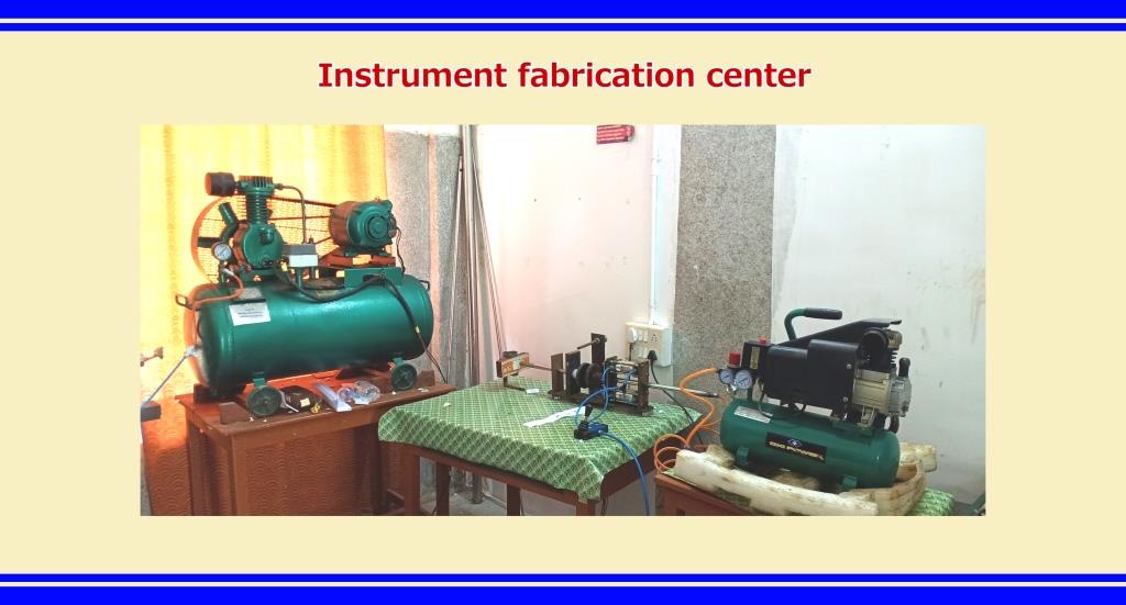
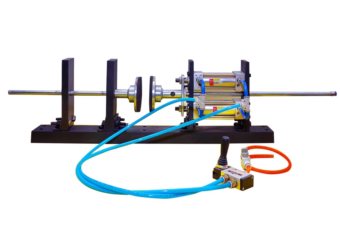
Table-top semi-automatic Reddy Tube is the product of Sacred Heart Instrumentation Centre, Sacred Heart College. This shock tube is capable of producing shock waves of Mach number ranging from 1 to 4.5. It has three sections as that of driver, driven and diaphragm sections. An air compressor is used to supply driver gas into the driven section. The driver and driven sections consist of long seamless stainless steel tubes of 48 cm and 180 cm, respectively and both have an inner diameter of 1.5 cm. While the atmospheric air is compressed into the driver section, at the critical pressure, the diaphragm is ruptured and the shock wave is generated and moves along the driven section. The sample holder is placed at 1 cm away from the open end of the driven section. The shock tube is capable to produce up to 500 shocks per hour as well as we can vary the Mach number by suitable diaphragms. More than 20 institutions including research laboratories from abroad used this shock tube and made more than 75 research publications in reputed international journals.
Recent progress in X-ray crystallography and in particular cryo-EM (cryo-electron microscopy) has greatly advanced our
understanding of the structure and dynamics of biomolecules. Although structures of several proteins have been solved in
different conformational states, the dynamics and the functional mechanism of how these macromolecules operate cannot be
understood from static snapshots obtained experimentally. Molecular dynamics (MD) simulations can be used to understand
the dynamic nature of biomolecular systems with high spatial and temporal resolution. The lab utilizes both coarse-grained models
and all-atom MD simulations to understand conformational dynamics in proteins and protein folding.
Our research interest includes understanding the conformational changes and dynamics of periplasmic binding proteins.
We also probe for structural features that guide the mechanism of ligand binding in these proteins.
The lab also studies ligand transport mechanism in membrane proteins, particularly glucose transporters and ATP-binding cassette (ABC) transporters.
The lab also focuses on understanding protein folding and domain-swapping. Our attention here is focused on probing the folding mechanism of proteins,
particularly those that domain-swap and relate this to understand the crucial mechanism that triggers folding as a domain-swapped dimer as against a monomer.
Lab facility
The lab is well equipped with 6 GPU and 6 high-end workstations, network access storage, printer, separate internet connection.
The lab has close collaborations with NCBS (Bengaluru) and IIT-Madras.
Research funding
1. Teacher Associateship for Research Excellence (TARE) award (Science & Engineering Research Board (SERB), Govt. of India), in collaboration with Dr. Athi Naganathan (Dept. of Biotechnology, IIT-Madras)
Duration: Three years (Dec. 2020-Nov. 2023)
Title:“Understanding the functional dynamics and drug efflux mechanism of ABC transporters”
Funds Sanctioned: Rs. 8,25,000
2. Early Career Research (ECR) award (SERB, Govt. of India), Apr. 2017-Mar. 2020 @ Sacred Heart College, Tirupattur
Duration: Three years (Mar. 2017-Mar. 2020)
Title:“Multiscale Modelling to Gain Mechanistic Insights into Glucose Transporters”
Funds Utilized: Rs. 18,90,010
